|
Anterior olfactory nucleus
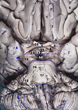
The anterior olfactory nucleus (AON; also called the anterior olfactory cortex) is a portion of the forebrain of vertebrates. It is involved in olfaction[1] and has supposedly strong influence on other olfactory areas like the olfactory bulb and the piriform cortex.[2] StructureThe AON is found behind the olfactory bulb and in front of the piriform cortex (laterally) and olfactory tubercle (medially) in a region known as the olfactory peduncle[3] or retrobulbar area. The peduncle contains the AON as well as two other much smaller regions, the taenia tecta (or dorsal hippocampal rudiment) and the dorsal peduncular cortex. FunctionThe AON plays a pivotal but relatively poorly understood role in the processing of odor information. Odors enter the nose (or olfactory rosette in fishes) and interact with the cilia of olfactory receptor neurons. The information is sent via the olfactory nerve (Cranial Nerve I) to the olfactory bulb. After the processing in the bulb the signal is transmitted caudally via the axons of mitral and tufted cells in the lateral olfactory tract. The tract forms on the ventrolateral surface of the brain and passes through the AON, continuing on to run the length of the piriform cortex, while synapsing in both regions. The AON distributes the information to the contralateral olfactory bulb and piriform cortex as well as engaging in reciprocal interactions with the ipsilateral bulb and cortex. Therefore, the AON is positioned to regulate information flow between nearly every region where odor information processing occurs. ComponentsThe AON is composed of two separate structures:
References
External links
|
||||||||||||||||||||||
